Enhancing pediatric safety: using simulation to assess radiology resident preparedness for anaphylaxis from intravenous contrast media

Tags

,

Radiology. 2007 Oct;245(1):236-44.

Enhancing pediatric safety: using simulation to assess radiology resident preparedness for anaphylaxis from intravenous contrast media.

Gaca AM1, Frush DPHohenhaus SMLuo XAncarana APickles AFrush KS.


PURPOSE:

To prospectively develop and test a simulation model for assessing radiology resident preparedness for pediatric life-threatening events in the radiology environment.

MATERIALS AND METHODS:

This study was institutional review board approved. Nineteen radiology residents (10 men, nine women; mean age, 28.5 years) participated in two simulated contrast material reaction scenarios: one with and one without resuscitation aids available. Each resident examined and managed two mannequins-simulating a 1-2-year-old patient and an 8-9-year-old patient-for type, sequence, dose, and administration route for any intervention, including administering medication, calling a code team, and providing oxygen. The time to order each intervention was documented. Resident responses (time to order intervention, appropriateness of intervention, and intervention route) were evaluated. The paired t test was used to compare the time to intervention between the resuscitation-aid-available and resuscitation-aid-not-available scenarios and between the scenario performed first and the scenario performed second. The McNemar test was performed to compare the percentage of appropriate interventions between the two resuscitation aid scenarios.

RESULTS:

The average time to call the code team was shorter when no resuscitation aids were available than when resuscitation aids were available (98 vs 149 seconds, P=.08). The average times to request oxygen and epinephrine were shorter when resuscitation aids were available (40 vs 89 seconds to request oxygen, P=.016; 121 vs 163 seconds to request epinephrine, P=.21). Appropriate medication dosing was not significantly different between the two scenarios. In only five of the 38 simulated scenarios was calling the code team the first intervention. The correct sequence of interventions (calling code team, providing oxygen, and then providing epinephrine) was performed by only one resident in one scenario. Only five residents recognized that they were encountering a contrast material reaction.

CONCLUSION:

Simulation training for radiology residents is valuable and suggests that resident preparedness for pediatric anaphylaxis from intravenous contrast media is insufficient. Clear step-by-step resuscitation aids are needed in the radiology environment.

FullSizeRender

Key Criteria for Selection of Radiology Residents: Results of a National Survey

Tags

Acad Radiol. 2006 Sep;13(9):1155-64.

Key criteria for selection of radiology residents: results of a national survey.


FullSizeRender

RATIONALE AND OBJECTIVES:

We sought to identify the criteria that academic radiology departments in the United States consider for selecting their residents.

MATERIALS AND METHODS:

In a cross-sectional study, a validated survey was sent to all the program directors of radiology residency programs. A total of 25 variables were studied. Descriptive statistics and correlations were calculated by the chi2 test. Nonparametric correlations were calculated with the Kruskal-Wallis rank test. Statistical significance was set at 5% alpha-error level (P<.05).

RESULTS:

We had a response rate of 53.1% (77 of 145). All responders participate in the National Resident Matching Program (NRMP), and 93.5% fill all their positions through NRMP. The preinterview selection criteria showed no significant difference by size, region, or affiliation with a medical school. An “interviewing body” carries out the interview process in 87.3% of the cases. Residents and fellows are part of the interviewing body in 76.5% of the programs, the body has the final word in accepting candidates in 62.9% of the programs, 55.4% of the programs use score sheets during interviews with candidates, and only 6.5% of the programs perform panel interviews. Programs associated with a medical school are significantly more likely to have more members in their interviewing body and to use score sheets when evaluating candidates, and panel interviews (more than one candidate or interviewer) are significantly more common among programs in the northeast region.

CONCLUSION:

All preinterview selection criteria and some interview structural characteristics are independent of the program’s size, region, or affiliation with a medical school. More research regarding optimal preselection and interview processes is needed, and closer attention should be paid to the NRMP process if current practices are to be maintained

Acute contrast reaction management by radiologists: A local audit study

Tags

, ,

  • Bartlett MJ, Bynevelt M. Acute contrast reaction management by radiologists: a local audit study. Australasian Radiology 2003;47:363– 367.

    (Department of Diagnostic Imaging and Interventional Radiology, Royal Perth Hospital, Perth, Western Australia, Australia)


  • Consultant radiologists & trainees must possess knowledge of optimal acute management of contrast reactions because patient survival depends upon prompt initial management

  • Questionnaire
    1. What is the emergency telephone number at your institution to summon help in an emergency 7700
    2. Name the initial dose of Adrenaline
      1. Initial doses of adrenaline (1:1000) up to 0.75mg (Adult); Intramuscular is the preferred route of administration
    3. Name an Antihistamine and its dose
      1. Promethazine (Antihistamine) 0.5~1mg/kg
    4. Name a Corticosteroid and its dose
      1. Hydrocortisone 2~6mg/kg OR Dexamethasone 0.1~0.4mg/kg
    5. Name the dose of Atropine for profound bradycardia
      1. 0.3~0.6 mg
    6. What additional route for drug administration can be used in paediatric patients?
      1. Intraosseous route for drug & fluid administration
    7. What is the dose in joules for cardioversion of Ventricular Fibrillation?
      1. 200~360 J
    8. What are the rates of cardiac massage/minute for inadequate cardiac output?A. Cardiopulmonary Resuscitation (CPR): A universal compression to ventilation ratio of 30:2 is recommended.   With children, if at least 2 trained rescuers are present a ratio of 15:2 is preferred. In newborns a rate of 3:1 is recommended unless a cardiac cause is known in which case a 15:2 ratio is reasonable.
    9. Name a type of intravenous fluid for volume expansion in the hypotensive patient
      1. Normal saline
    10. Name the initial dose of Adrenaline for a paediatric patient.
      1. 10micrograms/kg for children
  • Anaphylaxis/anaphylactoid reactions are severe systemic allergic reactions that include one or both of two severe features: respiratory difficulty& hypotension.
  • The most important single medication in the treatment of anaphylactic/anaphylactoid reaction is adrenaline.
    • It is sympathomimetic leading to peripheral vasoconstriction, increased cardiac contractility & bronchodilation
  • Less-acute management, including corticosteroid & anti-histamine doses, were assessed

Image guided percutaneous renal biopsy

Image guided percutaneous renal biopsy, utilising either ultrasound or CT allows for an accurate, reliable method of acquiring renal tissue for histopathological assessment.

Biopsy make be of a native or transplant kidney. It is divided into two types:

  • non-focal or non-targeted
  • focal or targeted (i.e. directed at a lesion)

Either type may be performed as a CT guided biopsy or as an ultrasound guided biopsy 1. Recent description has been given of the use of 3D cone beam CT in assisting the biopsy of particularly challenging focal lesions 2.

This depends on both patient and operator factors, such as patient body habitus, ability to cooperate and operator experience. Transplant renal biopsy is usually undertaken with ultrasound guidance given its more superficial location in the pelvis.

An alternative option for percutaneous CT/US guidance is the transjugular renal biopsy.

Continue p. 217

Ground glass opacity

Tags

, , ,

Screen Shot 2014-12-03 at 7.43.39 am Neumonitis_por_hipersensibilidad_2

  • Nonspecific finding on CT scans that indicates a partial filling of air spaces by exudate or transudate, as well as interstitial thickening or partial collapse of lung alveoli
  • DDx:
    • Pulmonary oedema
    • Infections (Cytomegalovirus & Pneumocystis carinii pneumonia)
    • Various non-infection: Interstitial lung diseases (e.g. hypersensitivity pneumonitis, Hamman-Rich syndrome)
    • Diffuse alveolar haemorrhage
    • Crytogenic organizing pneumonia

Screen Shot 2014-12-03 at 8.21.54 am

Response Evaluation Criteria In Solid Tumours (RECIST)

Tags

,

  • A set of published rules/criteria that define when tumours in cancer patients improve (respond), stay the same (stabilise) or worsen (progress) during treatments.
  • Published in Feb 2000 (updated in 2009) by an international collaboration including
    • European Organisation for Research & Treatment of Cancer (EORTC)
    • National Cancer Institute of the United States
    • National Cancer Institute of Canada Clinical Trials Group
  • Today, the majority of clinical trials evaluation cancer treatments for objective response in solid tumours are using RECIST
  • Specifically not meant to determine whether patients have improved or not, as these are tumour-centric, not patient centric criteria
    • It is not intended that these RECIST guidelines play a role in that decision making, except if determined appropriate by the treating oncologist
  • Can be used with CT, MRI or conventional radiography
  • Characterise lesions as measurable or non-measurable & target vs non-target

Anatomy of the adrenal (suprarenal) gland

Tags

, ,

Radiology & adrenal gland

  • Plain radiography, ultrasound, CT & MRI
    • The normal adrenal is never seen on plain films
    • On u/s it is easily seen in neonates (because it is larger) but rarely in adults
    • It has a thin reflective core, with a transonic outer portion
    • On CT & MRI,  the adrenals are almost always seen but are clearest in those with sufficient retroperitoneal fat

FullSizeRender

FullSizeRender

Nuclear medicine

  • Various scintigraphic methods & agents are used to provide metabolic information
  • MIBG (analogue of guanethidine) is used to image medullary disorders (such as phaeochromocytoma) as it is concentrated in sympathoadrenal tissue
  • Positron emission tomography (PET) with fluorine-18 (F-18) fluoro-deoxyglucose (FDG) can be used to differentiate between benign & malignant adrenal lesions depending on the degree of FDG uptake

The adrenal gland

  • Paired retroperitoneal glands, supero-medial to the kidneys within the perinephric space, but outside the renal capsule.
  • Develop between weeks 4 & 8 gestation
  • Endocrine organs with an:
    • Outer cortex of mesodermal origin
      • 3 Zones – zona glomerulosa, zona fasciculata & zona reticularis, that are fully differentiated by the 3rd year of life
      • Lipid-rich & secretes corticosteroids (aldosterone) & androgens
    • Inner medulla
      • Derived from neural crest cells, that migrate & inter-digitate into developing renal cortex
      • Darker colour
      • Related to the sympathetic nervous system & secrete catecholamines
    • At birth, the cortex is much larger & later regresses. The adrenal glands are 1/3 the weight of the adjacent kidney at birth, but 1/30 in the adult
    • Adult dimensions
      • Cranio-caudal length 2~4cm
      • Body R) 4~8mm, L) 6~10mm
      • Width of limbs 3~4mm
    • Variable shape
      • R) gland is linear or inverted ‘V shape
      • L) gland is inverted ‘Y’ or ‘V’

FullSizeRender

Neurovascular & lymphatic anatomy

  • 3 arteries on each side:
    • Superior adrenal artery from the inferior phrenic artery (which is a branch of the abdominal aorta)
    • Middle adrenal artery arises from the abdominal aorta
    • Inferior adrenal artery from the renal artery
  • A single vein drains each gland:
    • The shorter R) adrenal vein drains directly into the IVC
    • The longer L) adrenal vein empties into the L) renal vein & may be joined by the inferior phrenic vein
  • Neural anatomy:
    • Preganglionic sympathetic fibres from the splanchnic nerves
    • Postganglionic vasomotor fibres are distributed with the arteries supplying the gland to regulate its blood flow

FullSizeRender

Normal variants of the adrenal glands

  • Unilateral absent glands are unknown
  • Ectopic adrenal cortical tissue may occur nearby (usually around the coeliac axis) but may also be displaced with the descending gonads during embryolo   gical development, and be found in the broad ligament, spermatic cord, testis or epididymis.
    • Ectopic adrenal tissue is present up to 50% of neonates, but only 1% adults as it involutes.
    • More distal ectopic adrenal tissue contains cortex alone

Port-a-Cath

Tags

,

800px-PAC_met_Gripper_erin

Definition:

  • A small medical appliance that is installed beneath the skin
  • A catheter connects the port to a vein
  • Under the skin, the port has a septum through which drugs can be injected & blood samples can be drawn many times, usually with less discomfort for the patient than a more typical “needle stick”
  • Used mostly to treat
    • Haematology & oncology patients
    • Haemodialysis patients
  • Usually inserted in the upper chest, just below the clavicle, leaving he patient’s hands free
  • The main form of a implantable central venous catheter or line

Terminology:

  • A portmanteau of “portal” & “cathether”
  • Port-a-Cath is a registered trade mark of Smiths Medical
  • The term totally implantable venous access system (TIVAS) is also used

How it works:

  • The catheter runs from the portal and is surgically inserted into a vein (usually the jugular vein, subclavian vein, or superior vena cava).
    • Ideally, the catheter terminates in the SVC, just upstream of the right atrium. This position allows infused agents to be spread throughout the body quickly & efficiently

Gray576

Risks:

  1. Infection
  2. Thrombosis – to prevent clotting the portacath is flushed with saline & heparin
  3. Mechanical failure
  4. Age
  5. Pneumothorax
  6. Arterial injury

Portkatheter_Röntgen

NB:

CVC can be divided into 

  1. Peripherally inserted central catheters (PICC)
  2. Non-tunnelled CVCs
    1. Used in ICU or ED for emergent or short term access  (<7~10 days)
    2. Vascath used for haemodialysis
  3. Tunnelled CVCs
    1. Hickman’s catheters, permacath
  4. Implantable ports
    1. e.g. Port-a-Cath, Infus-a-Port
    2. May be located in the chest or arm (brachial)
    3. May be single or dual lumen

MCQ Radiology Anatomy – Chest & Cardiovascular

Tags

,

1. Regarding the imaging modalities of the chest:

a) HRCT uses 1-2mm slice thickness & a high resolution computer algorith to show fine detail of the lung parenchyma, pleura and tracheobronchial tree. It is not used to delineate mases in the lung.

b) Spiral CT ensures that no portion of the chest is missed due to variable inspiratory effort.

c) Currently MRI is a poor technique for showing lung detail. It allows visualisation of the chest wall, heart, mediastinal and hilar structures.

d) Bronchoscopy is invasive technique, largely been superseded by HRCT.

e) CTPA is performed to diagnose majory pulmonary emboli using a cannula placed in any peripheral vein & is relatively non-invasive compared to conventional pulmonary angiography.


2. Regarding the development of the lung:

a) The tracheobronchial groove appears on the ventral aspect of the caudal end of the pharynx

b) The primary bronchial buds develop from the tracheobronchial diverticulum; The bronchial buds differentiate into bronchi in each lung

c) During embryonic life that alveoli is lined by cuboidal epithelium that lines the rest of the respiratory tract. When respiration commences at birth the transfer to the flattened pavement epithelium of the alveoli is accomplished.

d) Tracheo-oesophageal fistula (TOF) indicates the close developmental relationship between the foregut & the respiratory passages. It is usually associated with an atresia of the oesophagus & the fistula situated below the atretic segment

e) Uni-lateral pulmonary hypoplasia is usually due to congenital diaphragmatic hernia


3. Regarding the blood supply to the chest wall:

a) There are usually 9 pairs of posterior ateries from the postero-lateral margin of the thoracic aorta, distributed to the lower 9 intercostal spaces. The first and second spaces are supplied by the superior intercostal artery, branches of the costocervical trunk from the subclavian artery.

b) The internal thoracic artery arises from the subclavian artery & supplies the upper 6 intercostal spaces.

c) The neurovascular bundle passes around the chest wall in the subcostal groove deep to the internal intercostal muscle.

d) The intercostal spaces are drained by 2 anterior veins & a single posterior intercostal vein.

e) Posterior intercostal veins drain into the brachiocephalic vein & azygos system.  The anterior veins drain into the musculo-phrenic & internal thoracic veins.


4. Regarding the azygos venous system:

a) The azygos vein at the level of the 4th thoracic vertebra arches over the root of the right lung to end in the superior vena cava (SVC)

b) In 1% of the population, the azygos vein traverses the lung before entering the SVC resulting in the azygos fissure. The azygos ‘lobe’ is not a true segment.

c) The thoracic duct & aorta are to the left of the azygos vein

d) The 2nd, 3rd and 4th intercostal spaces on the right, drain via the right superior intercostal vein into the azygos vein; Hemiazygos, accessory hemiazygos, oesophageal, mediastinal, pericardial & right bronchial veins drain into the azygos system

e) In congenital absence of the IVC the azygos vein enlarges; In the azygos continuation of the IVC, the azygos is a large structure, but otherwise the anatomy is unaltered. This may be confused with a mediastinal mass.


5. Regarding the hemiazygos & accessory hemiazygos venous systems:

a) The hemiazygos vein at the level of the 8th thoracic vertebra crosses the vertebral column behind the aorta, oesophagus & thoracic duct.

b)

c)

d)

e)


6. Regarding the airways:

a)

b)

c)

d)

e)

MCQs AIT

Tags

,

1. Regarding atomic structure:

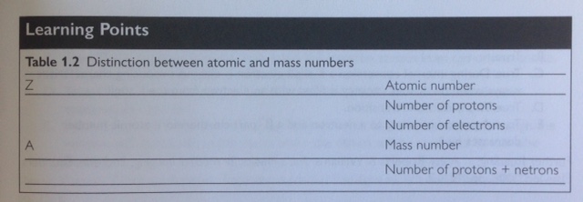
A. Z is the atomic number & indicates the number of protons in the nucleus

B. A is the mass number. Z determines place in the periodic table

C. Higher atomic number nuclei require more neutrons than protons for stability

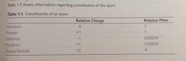
D. Neutrons have no charge, protons +1

E. They (Protons to neutrons in the nucleus) are tightly bound


2. Concerning orbital electrons

A. Electrons are arranged in shells around the nucleus at specific energy levels

B. Binding energy is expended completely removing the electron from the atom

C. It (Binding energy) is lowest for valence shell electrons and highest for the K shell

D. Characteristic radiation is from inner shells. The valence shell gives the chemical properties

E. K shell binding energy increases with increasing atomic number


3. Regarding the structure of atoms:

A. A proton has a mass approximately 1850x that of an electron

B. An electron is not a nucleon; Neutrons & Protons are nucleons

C. Positrons have the same mass as electrons

D. Alpha particles consists of 2 protons & 2 neutrons (helium nucleus), therefore the mass is 4x that a proton

E. There can be up to 2 electrons in the K shell, 8 in the L shell, 18 in the M shell and 32 in the N shell

FullSizeRender


4. Nuclides:

A. Each particular combination of Z and A defines a nuclide

B. Nuclides with the same number of protons but different number of neutrons are isotopes, therefore they have the same atomic numbers and different atomic mass

C. Not all are radioactive

D. Have the same chemical properties between isotopes of a particular element; Isotopes have the same number of protons and therefore when neutral the same number of electrons

E. May emit radiation if they have too few or too many neutrons

FullSizeRender


5. Regarding the electromagnetic spectrum

A. Sound waves do no fall within the spectrum

B. Frequency increases with energy, but velocity is constant

C. Frequency and wavelength of electromagnetic radiation are directly proportional to each other

D. All electromagnetic radiation travels at the speed of light in a vacuum; In a vacuum. velocity of radio waves is equal to that of infrared light.

E. UV light is further up the spectrum, therefore has a shorter wavelength than visible light.


6. Electromagnetic radiation:

A. Travels in straight lines if unattenuated

B. Have wave- & particle-like properties

C. Has energy that is usually expressed in electron volts (eV), which give manageable numbers for calculations (1eV = 1.6 x 10^-19Joules)

D. Comprises sinusoidally varying electric and magnetic fields perpendicular to each other & to the direction of travel

E. Does not include beta radiation; Beta particles are electrons emitted from the nucleus


7. Ionizing radiation:

A. Causes direct damage if it is absorbed in tissue

B. Causes indirect damage through ionisation of water & production of free radicals

C. Does not always obey the inverse square law; Only applicable to types of electromagnetic radiation from a point source and without attenuation

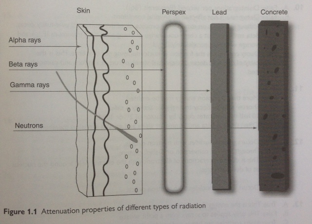
D. Gamma & X-rays are useful, neutrons, alpha, and beta particles are not

E. Beta radiation may require only Perspex shielding, however optimal shielding is achieved with Perspex backed with lead

FullSizeRender


8. Regarding secondary electrons:

A. They are recoil electrons produced during Compton scattering events

B. Their range depends upon the density of the material through which they are travelling & range also depends upon their initial energy

C. They interact with outer shell electrons to cause ionisation

D. They are the reason that x-ray and gamma rays are indirectly ionising; Alpha & beta particles are directly ionising as they are charged

E. Energy from the x-ray beam is converted into increased molecular motion & therefore heat.


9. In radioactive decay:

A. A neutron converts to a proton & a beta- particle, therefore atomic number increases by 1

B. Alpha particles are helium nuclei

C. Some radionuclides emit electrons and characteristic x-rays; During internal conversion a K shell electron is ejected, producing characteristic x-rays when the K shell vacancy is filled with an electron from the L shell.

D. Most nuclides left in the metastable state after beta decay, emit gamma rays to reach ground state; This is isometric transition

E. Positron emission reduces the number of protons in an atom by 1; A proton converts to a neutron and a beta+ particle, therefore atomic number decreases by 1


10. Regarding radioactivity:

A. Radioactive decay is the number of disintegrations per second (Bequerels (Bq))

B. Decay rate is not affected by physical conditions

C. If stored long enough, the radioactivity of a radionuclide will drop to zero; Although decay is an exponential process, all of the atoms will eventually decay.

D. Beta emission is at a continuous range of energies up to a maximum (Emax), and with average energy approximately Emax/3

E. Decay constant is the fraction of nuclei decaying per unit time. This is the probability of decay, as decay of individual atoms occurs at random and cannot be predicted.


11. The following are true of radionuclides:

A. Physical half-life (t1/2) is the time taken for the activity to decay to 1/2 the original value

B. Gamma rays are emitted at more than 1 photon energy

C. Gamma emitting radionuclides with shorter t1/2 are safer to use and store than thos with longer t1/2; Shorter time to decay to negligible activity is safer

D. In 10 half-lives the activity is reduced by a factor of approximately 1000; 10 half lives indicates decay by a factor of 2^10 or 1024

E. Often there is beta & gamma, or alpha & gamma emission together


12. Direct emission from radioactive decay includes:

A. Beta minus emission; Occurs in radionuclides with neutron excess

B. Characteristic x-rays; Through internal conversion or K-shell capture

C. Bremsstrahlung is due to interactions of electrons with the electric field around the nucleus and not of decay directly.

D. Alpha particles

E. Positron emission; Occurs in radionuclides with neutron deficit


13. Concerning properties of x-rays:

A. Beam intenstiy is the total energy per unit area per unit time; This is the energy fluence rate or intensity

B. The inverse square law applies to x-rays from a point source

C. X-rays have lower linear energy transfer than alpha particles; Alpha particles are heavy & produce ionizing events closely spaced along a short path, causing maximum DNA damage.

D. Only high-energy photons (x-rays/ gamma rays) are ionizing

E. At equivalent energy, an x-ray cannot be distinguished from a gamma ray; How they are produced differs, but they are indistinguishable at equivalent energies.