
Tags
Radiology & adrenal gland
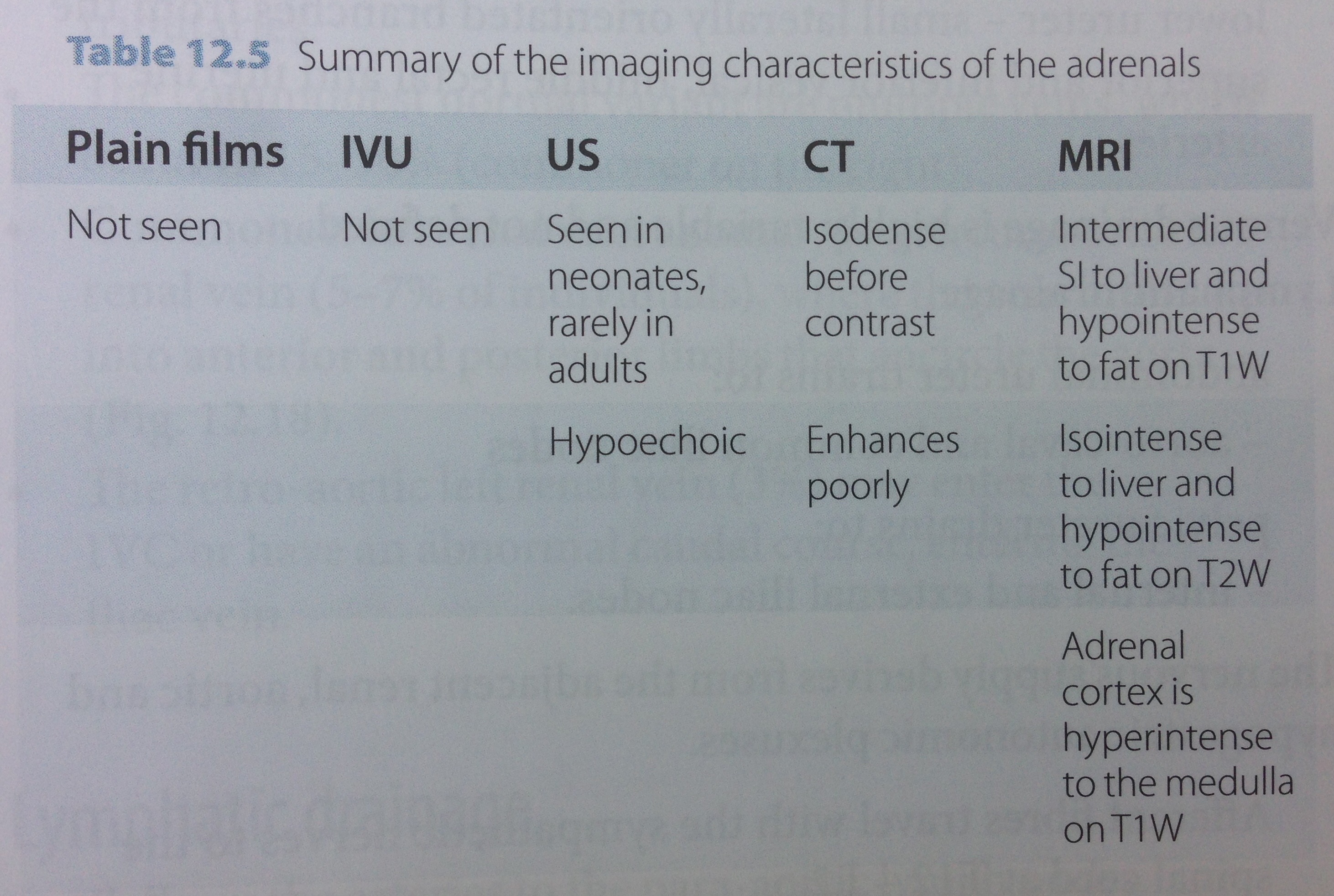
- Plain radiography, ultrasound, CT & MRI
- The normal adrenal is never seen on plain films
- On u/s it is easily seen in neonates (because it is larger) but rarely in adults
- It has a thin reflective core, with a transonic outer portion
- On CT & MRI, the adrenals are almost always seen but are clearest in those with sufficient retroperitoneal fat

Nuclear medicine
- Various scintigraphic methods & agents are used to provide metabolic information
- MIBG (analogue of guanethidine) is used to image medullary disorders (such as phaeochromocytoma) as it is concentrated in sympathoadrenal tissue
- Positron emission tomography (PET) with fluorine-18 (F-18) fluoro-deoxyglucose (FDG) can be used to differentiate between benign & malignant adrenal lesions depending on the degree of FDG uptake
The adrenal gland
- Paired retroperitoneal glands, supero-medial to the kidneys within the perinephric space, but outside the renal capsule.
- Develop between weeks 4 & 8 gestation
- Endocrine organs with an:
- Outer cortex of mesodermal origin
- 3 Zones – zona glomerulosa, zona fasciculata & zona reticularis, that are fully differentiated by the 3rd year of life
- Lipid-rich & secretes corticosteroids (aldosterone) & androgens
- Inner medulla
- Derived from neural crest cells, that migrate & inter-digitate into developing renal cortex
- Darker colour
- Related to the sympathetic nervous system & secrete catecholamines
- At birth, the cortex is much larger & later regresses. The adrenal glands are 1/3 the weight of the adjacent kidney at birth, but 1/30 in the adult
- Adult dimensions
- Cranio-caudal length 2~4cm
- Body R) 4~8mm, L) 6~10mm
- Width of limbs 3~4mm
- Variable shape
- R) gland is linear or inverted ‘V shape
- L) gland is inverted ‘Y’ or ‘V’
- Outer cortex of mesodermal origin
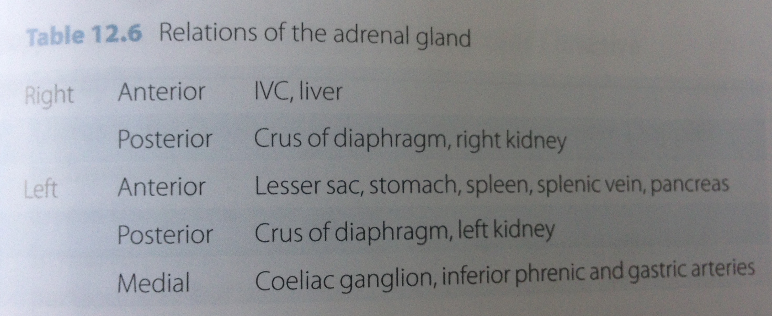
Neurovascular & lymphatic anatomy
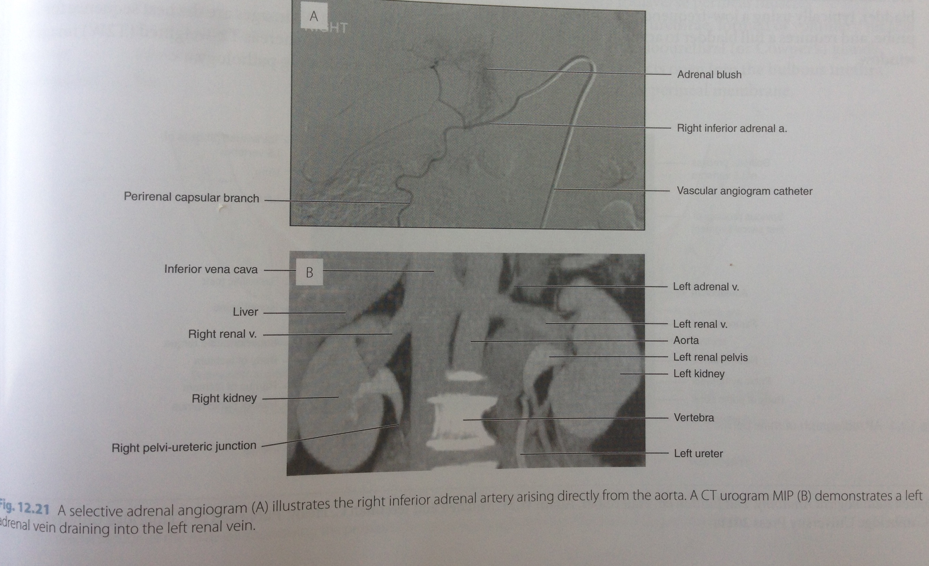
- 3 arteries on each side:
- Superior adrenal artery from the inferior phrenic artery (which is a branch of the abdominal aorta)
- Middle adrenal artery arises from the abdominal aorta
- Inferior adrenal artery from the renal artery
- A single vein drains each gland:
- The shorter R) adrenal vein drains directly into the IVC
- The longer L) adrenal vein empties into the L) renal vein & may be joined by the inferior phrenic vein
- Neural anatomy:
- Preganglionic sympathetic fibres from the splanchnic nerves
- Postganglionic vasomotor fibres are distributed with the arteries supplying the gland to regulate its blood flow
Normal variants of the adrenal glands
- Unilateral absent glands are unknown
- Ectopic adrenal cortical tissue may occur nearby (usually around the coeliac axis) but may also be displaced with the descending gonads during embryolo gical development, and be found in the broad ligament, spermatic cord, testis or epididymis.
- Ectopic adrenal tissue is present up to 50% of neonates, but only 1% adults as it involutes.
- More distal ectopic adrenal tissue contains cortex alone
